Microscopy & Histology Core Facility
Overview Head of the Core Facility Core Facility Members Publications Terms of useThe Microscopy and Histology Core Facility offers a comprehensive array of high-performance microscopes and expert support to ensure top-notch and reliable imaging.
The Microscopy and Histology Core Facility provides you with hands-on training and access to state-of-the-art instruments to visualise both living and fixed cells. Our complete instrument line-up can be found on OpenIRIS, our booking software.
We also offer tissue processing, embedding and sectioning, vibratome sectioning and cryo-sectioning devices, as well as cell culture facilities for sample preparation. For histology, we provide support with machines and basic protocols (e.g. H&E staining, in situ hybridisation, immunostaining).
Our facility staff have a broad range of research experience and technical expertise. We are happy to provide feedback on your experimental design and assist with troubleshooting and method development at any time.
To use our services, please get in touch (microscopy(@)imb-mainz.de) to schedule a mandatory introductory session and user meeting discussing your project needs, biosafety and our user guidelines. Researchers from IMB, Mainz University, its University Medical Center and the local Max Planck Institutes are eligible to use our facility. Access for non-associated researchers is decided on a case-by-case basis.
Full service:
Full experiment design, imaging, processing, analysis and figure preparation
Self services:
- Independent booking and use of microscopes (after training)
- Independent booking and use of histology instruments (after training)
Support services:
- Comprehensive training for independent use of instruments
- Assistance with planning and designing experiments
- First level support and ad hoc troubleshooting during working hours
- Assistance with image analysis
- Proofreading of methods sections for publications
Key instruments
Widefield
AF7000 Widefield Fluorescence Microscope - live (Room 00.431)
The AF7000 (Leica) is an inverted widefield microscope with fluorescence (UV, green, red and far red fluorescence) and diverse contrast methods in transmitted light (brightfield, phase contrast, DIC, polarisation). It is equipped with an environmental control box (temperature, CO2) for live cell imaging and a software module for small screening applications.
- Available objectives: 1.25x/0.04 air, 10x/0.3 air, 20x/0.4 air or 20x/0.8 air, 40x/0.6 air, 63x/1.4 oil, 63x/1.2 water, 100x/1.4 oil
Thunder Widefield Fluorescence Microscope - live (Room -1.291)
The THUNDER (Leica) is an inverted widefield microscope with fluorescence and built-in image processing tools for background subtraction and adaptive deconvolution (based on Leica LIGHTNING).
Diverse contrast methods in transmitted light (brightfield, phase contrast, DIC) and a color camera are available. It is equipped with an environmental control box (temperature, CO2) for live cell imaging and a software module for small screening applications.
- Available objectives: 10x/0.3 dry, 25x/0.95 water (long working distance 2.5 mm), 40x/0.95 air, 63x/1.4 oil, 100x/1.44 oil
- 8 LEDs: 395, 438, 475, 511, 555, 575, 635 and 730 nm
DeltaVision Widefield Fluorescence Microscope - live (Room 00.431)
Specialized widefield microscope (Leica) for imaging yeast, with the possibility to perform deconvolution directly on the system. Incubation chamber with heating and cooling option.
Spinning disk
VisiScope Spinning Disk Confocal Microscope – live (Room -1.261)
The Spinning Disc Confocal (5 Elements, Visitron) microscope has an ablation laser on a Nikon stand with a CSU-W1 spinning disk (25 and 50 µm pinholes) from Yokogawa. It is equipped with two sCMOS cameras for simultaneous live cell imaging in two channels and seven laser lines. The microscope is optimised for live cell imaging with TIRF, FRAP and DNA ablation options (355 nm pulsed laser).
- Available objectives: 10x/0.45 air, 10x/0.5 gly/water/oil, 20x/0.75 air, 40x/0.95 air, 20x/0.95 water, 25x/1.05 silicone, 40x/1.25 water, 60x/1.2 water, 100x/1.49 oil
BC43Spinning Disk Confocal Microscope – live (Room 00.413)
The BC43 (Andor) is a prime solution for 3D imaging thick samples. A high-sensitivity, high-speed imaging camera makes it perfect for live-cell confocal imaging. Widefield and transmitted light modes are available as well. A motorised XY stage allows multiple-position acquisition, large organism montages and multipoint scanning, resulting in outstanding productivity from a single imaging experiment.
- Epi and confocal fluorescence mode
- Four excitation lines (405, 488, 561, 640nm)
- Available objectives: 2x air, 10x/0.45 air, 20x/0.75 air, 40x/0.75 air, 60x (1.4) oil
Point-scanning confocal/STED
Stellaris 8 FALCON Confocal Laser Scanning Microscope with Fluorescence Lifetime Option (Room 00.421)
The Stellaris 8 FALCON (Leica) is an inverse confocal fluorescence scanning microscope with the capability to perform Fluorescence Lifetime Imaging Microscopy (FLIM). It is optimal for imaging living and fixed samples with multicolor fluorescence.
- FLIM can be used for several applications, such as improving image quality by removing autofluorescence, multiplexing fluorophores with overlapping spectra, detecting cellular metabolites based on their fluorescence lifetime, and measuring molecular interactions with FRET sensors
- Deconvolution module (Lightning)
- FRAP module
- Fast scanning with a resonance scanner
- Equipped with a pulsed white light laser (excitation range 440-790 nm, 80MHz pulse frequency) and diode lasers (405 nm, 488 nm, 561 nm)
- The fluorescence detection range can be continuously adjusted between 400 - 850 nm and emission is detected by optimized photomultipliers (HyD-S, HyD-X, HyD-R)
- Mosaic scanning of large objects is possible due to the motorised xy-stage
- Available objectives: 10x/0.4 air, 20x/0.75 air, 40x/1.3 oil, 63x/1.4 oil, 100x/1.4 oil, 63x/1.2 water
SP5 Confocal Laser Scanning Microscope (Room 00.461)
The TCS SP5 (Leica) is an inverse confocal fluorescence microscope optimised for imaging fixed samples with multicolour fluorescence. Four lasers are available for excitation (405, argon (458, 476, 488, 495, 514), 561 and 633 nm). The fluorescence detection range can be continuously adjusted between 400 - 800 nm and emission is detected by 4 PMTs. Mosaic scanning of large objects is possible due to the motorised xy-table.
- Available objectives: 10x/0.3 dry, 20x/0.7 dry, 40x/1.3 oil, 63x/1.2 water, 63x/1.4 oil
SP5 STED CW Super-Resolution Microscope (Room -1.261)
The TCS SP5 STED CW (Leica) confocal microscope is dedicated to super-resolution microscopy based on STED (STimulated Emission Depletion) and live cell imaging. Super-resolution in the xy-direction (ca. 70 nm) is achieved for fluorophores with emission spectra overlapping with the 592 nm depletion laser. Additionally, the setup is equipped with an FCS module (Picoquant) and APD sensors to measure diffusion rates. Three lasers with the following wavelengths are available for excitation: argon (458, 476, 488, 495, 514), 561 and 633 nm. The detection range can be continuously adjusted between 400 - 800 nm and emission is detected by 3 PMTs and 2 HyDs.
- Available objectives: 10x/0.3 dry, 20x/0.7 water/oil, 40x/1.1 water, 63x/1.2 water, 63x/1.4 oil, 100x/1.4 oil
Holotomography
CX-F 3D Cell Explorer - live (Room -1.291)
The 3D Cell Explorer fluo (Nanolive) is a holo-tomographic widefield microscope with fluorescence options for gentle live cell imaging. The contrast is based on refractive index measurements in living samples and provides "label-free" discrimination of several organelles (mitochondria, lipid vesicles, nuclei, nucleoli, lysosomes, etc.).
Whole 3D volumes as large as 80 µm x 80 µm x 30 µm can be imaged within 2 seconds. The set-up is equipped with a 60x air objective and an incubation chamber (temperature, CO2 settings available) for live cell imaging. Fluorescence imaging options (widefield) for green, red and far-red fluorophores are available.
High-content screening
IncuCyte SX5 Widefield Fluorescence Screening Microscope - live (Room 00.281)
The IncuCyte SX5 (Sartorius) is a box-type widefield screening microscope for live cell imaging with several predefined assays and analysis pipelines. The setup is equipped with 4x, 10x and 20x air objectives and located inside an incubator chamber (temperature, CO2 settings available) ideally for long-term live cell imaging. There are six slots for multiwell plates (6, 12, 24, 48 or 96 well) and/or four slots for chambered slides.
Two fluorescence detection modules are available:
- PH/G/R: Phase contrast/green (441-481 nm, 503-544 nm)/red (567-607 nm, 622-704 nm)
- PH/G/O/NIR: Phase contrast/green (453-485 nm, 494-533 nm)/orange (546-568 nm, 576-639 nm)/NIR (648-674 nm, 685-756 nm)
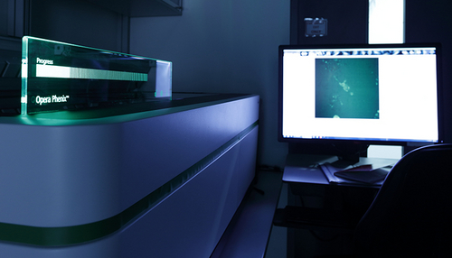
Opera Phenix Spinning Disk Confocal Microscope – live (Room 00.531)
The Opera Phenix (Revvity) is a high-content screening (HCS) microscope system, built to handle multi-well plates as well as slides. The system can be operated in widefield mode and spinning-disc confocal mode. Fixed and living cells/organisms can be recorded. The system is equipped with two sCMOS cameras for fast recording, with four laser lines (405, 488, 561 and 640 nm).
- Available objectives: 1.25x/0.03 air, 10x/0.3 air, 20x/1.0 water, 40x/1.1 water, 63x/1.15 water
Super-resolution/TIRF
NanoImager Super-Resolution Microscope (Room 00.413)
The NanoImager is a compact benchtop widefield microscope dedicated to single-molecule localization microscopy (SMLM). Resolution in the range of 20 nm in xy (STORM, DNA-PAINT, PALM) and 100 nm in Z (+/- 600 nm) can be obtained using this method. SMLM in multiple colors (four lasers) and two simultaneous channels (dichroic mirror split at 640 nm) is possible.
- Excitation laser power at sample: 405 nm (15 mW), 488 nm (250 mW), 561 nm (250 mW), 640 nm (250 mW)
- Excitation laser power at source: 405 nm (1 W), 488 nm (1 W), 561 nm (750 mW), 640 nm (1 W)
- Available objective: 100x/1.45 oil
- Motorised xyz stage
- TIRF/HiLo mode
GSDIM Super-Resolution Microscope (Room -1.261)
The GSD 3D (Leica) widefield microscope is dedicated to super-resolution microscopy based on fluorophore blinking achieved by GSD (Ground State Depletion) and the single-molecule localisation algorithm (also called dSTORM or PALM). Structures in the range of 40 nm (xy) and 50 nm (z) can be resolved. The microscope is further equipped with a TIRF (total internal reflection) module, which is ideal for imaging samples located close to the cover glass/water interface, such as membrane receptors. The objective for super-resolution and TIRF is 160x/1.47 oil.
SP5 STED CW Super-Resolution Microscope (Room -1.261)
The TCS SP5 STED CW (Leica) confocal microscope is dedicated to super-resolution microscopy based on STED (STimulated Emission Depletion) and live cell imaging. Super-resolution in the xy-direction (ca. 70 nm) is achieved for fluorophores with emission spectra overlapping with the 592 nm depletion laser. Additionally, the setup is equipped with an FCS module (Picoquant) and APD sensors to measure diffusion rates. Three lasers with the following wavelengths are available for excitation: argon (458, 476, 488, 495, 514), 561 and 633 nm. The detection range can be continuously adjusted between 400-800 nm and emission is detected by 3 PMTs and 2 HyDs.
- Available objectives: 10x/0.3 dry, 20x/0.7 water/oil, 40x/1.1 water, 63x/1.2 water, 63x/1.4 oil, 100x/1.4 oil
FRAP, FRET, FLIM, FCS, laser ablation
FRAP/TIRF/Ablation: VisiScope Spinning Disk Confocal Microscope – live (Room -1.261)
FRAP/FLIM: Stellaris 8 FALCON Confocal Laser Scanning Microscope with Fluorescence Lifetime Option (Room 00.421)
FRAP/FCS: SP5 STED CW Super-Resolution Microscope (Room -1.261)
Label-free imaging
CX-F 3D Cell Explorer - live (Room -1.291)
The 3D Cell Explorer fluo (Nanolive) is a holo-tomographic widefield microscope with fluorescence options for gentle live cell imaging. The contrast is based on refractive index measurements in living samples and provides "label-free" discrimination of several organelles (mitochondria, lipid vesicles, nuclei, nucleoli, lysosomes, etc.).
Whole 3D volumes as large as 80 µm x 80 µm x 30 µm can be imaged within 2 seconds. The set-up is equipped with a 60x air objective and an incubation chamber (temperature, CO2 settings available) for live cell imaging. Fluorescence imaging options (widefield) for green, red and far-red fluorophores are available.
Optical tweezer
Lumicks C-Trap (Room –1.281)
The C-Trap (Lumics) combines high-resolution optical tweezers with advanced microfluidics and multicolor confocal imaging, allowing manipulation and visualisation of single molecules and the measurement of piconewton forces. The system is designed for automated control over the flow of up to five solutions, with four optical traps and confocal imaging with three laser excitation lines (488, 561 and 638 nm).
Deconvolution
The Huygens Remote Manager provides access to web-based deconvolution by the Huygens Software (SVI). Here you can upload your images to the university server and start the deconvolution. You will receive an email notification when the deconvolution has finished.
Image data analysis & AI
Five image processing stations (Room 00.457)
Both commercial (Imaris, vision 4D, VisiView, LAS-X, Zen blue Intellesis, Huygens and Huygens Remote Manager, Harmony, Columbus, GraphPad Prism, MatLab) and open source (e.g. Fiji (ImageJ), QuPath, Icy) software tools are available, on-site and remotely.
RDM Research data management
Lectures and courses
We offer advanced training through practical courses and a variety of lectures. Practical courses focus on image processing and analysis (e.g. from basics to simple macro programming in Fiji, Icy, deconvolution with Huygens, Imaris) and super-resolution microscopy.
Microscopy lectures
- Introduction to microscopy
- F-techniques and super-resolution
- Histology and fluorescent labelling
- Image manipulation: the slippery slope to misconduct
Microscopy courses
Image processing and analysis:
- Basics of image analysis with Fiji/ImageJ, including filtering, segmentation, analysing objects, quantification, co-localisation, setting scale bars, visualisation of multidimensions, tracking
- 3D visualisation and analysis with Imaris and Ilastik (machine learning)
- Deconvolution with Huygens Remote Manager (HRM)
- Recording and writing macros in Fiji
- Ethics of image processing
Super-resolution microscopy:
- Introduces techniques to resolve fluorescent structures below the diffraction limit of light (ca. 250 nm in xy or 500 nm in z)
- Theoretical background of current techniques, sample preparation, hands-on STED (Stimulated Emission Depletion) and SMLM (Single Molecule Localization Microscopy), as well as data analysis
Acknowledgements/Co-authorships
“An acknowledgement of the Core Facilities in your publication is important and more than just a nice way to say thank you. Your acknowledgement matters!”
Acknowledgements are the currency by which Core Facilities are measured: they help us prove our value and our contribution to research results. Acknowledging Core Facilities shows that we are important partners in the scientific community, and it allows us to maintain funding and make further investment in the Core Facilities. In the case that Core Facility staff members have contributed significantly to your research project, we ask that you treat them the same way you would treat any other collaborator and consider a co-authorship.
In accordance with DFG recommendations, we furthermore ask that you acknowledge any instrumentation that is funded through a DFG major instrumentation grant by including the corresponding project number.
Communities
We are a member of German BioImaging (GerBI)

If you would like to get in touch with the local microscopy community alias Mainz Microscopy Connection (MMC), don´t hesitate to contact us or the MMC directly (mmc(@)imb.de).


Think global (https://microscopydb.io/who-we-are/#partners), image local.

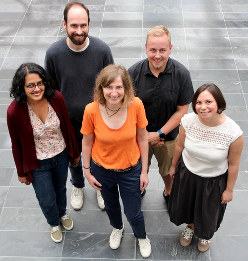
Sandra Ritz is a principal investigator and Anusha Bargavi Gopalan is a member of the SFB 1551 on “Polymer Concepts in Cellular Function”.